Rigid Flat Feet In Adults
Overview
One in four adults in the U.S. has adult acquired flatfoot deformity, which may begin during childhood or be acquired with age. The foot may be flat all the time or may lose its arch when the person stands. The most common and serious cause of flat foot is Posterior Tibial Tendon Dysfunction, in which the main tendon that supports the arch gradually weakens. 
Causes
There are multiple factors contributing to the development of this problem. Damage to the nerves, ligaments, and/or tendons of the foot can cause subluxation (partial dislocation) of the subtalar or talonavicular joints. Bone fracture is a possible cause. The resulting joint deformity from any of these problems can lead to adult-acquired flatfoot deformity. Dysfunction of the posterior tibial tendon has always been linked with adult-acquired flatfoot deformity (AAFD). The loss of active and passive pull of the tendon alters the normal biomechanics of the foot and ankle. The reasons for this can be many and varied as well. Diabetes, high blood pressure, and prolonged use of steroids are some of the more common causes of adult-acquired flatfoot deformity (AAFD) brought on by impairment of the posterior tibialis tendon. Overstretching or rupture of the tendon results in tendon and muscle imbalance in the foot leading to adult-acquired flatfoot deformity (AAFD). Rheumatoid arthritis is one of the more common causes. About half of all adults with this type of arthritis will develop adult flatfoot deformity over time. In such cases, the condition is gradual and progressive. Obesity has been linked with this condition. Loss of blood supply for any reason in the area of the posterior tibialis tendon is another factor. Other possible causes include bone fracture or dislocation, a torn or stretched tendon, or a neurologic condition causing weakness.
Symptoms
Symptoms shift around a bit, depending on what stage of PTTD you?re in. For instance, you?re likely to start off with tendonitis, or inflammation of the posterior tibial tendon. This will make the area around the inside of your ankle and possibly into your arch swollen, reddened, warm to the touch, and painful. Inflammation may actually last throughout the stages of PTTD. The ankle will also begin to roll towards the inside of the foot (pronate), your heel may tilt, and you may experience some pain in your leg (e.g. shin splints). As the condition progresses, the toes and foot begin to turn outward, so that when you look at your foot from the back (or have a friend look for you, because-hey-that can be kind of a difficult maneuver to pull off) more toes than usual will be visible on the outside (i.e. the side with the pinky toe). At this stage, the foot?s still going to be flexible, although it will likely have flattened somewhat due to the lack of support from the posterior tibial tendon. You may also find it difficult to stand on your toes. Finally, you may reach a stage in which your feet are inflexibly flat. At this point, you may experience pain below your ankle on the outside of your foot, and you might even develop arthritis in the ankle.
Diagnosis
Your podiatrist is very familiar with tendons that have just about had enough, and will likely be able to diagnose this condition by performing a physical exam of your foot. He or she will probably examine the area visually and by feel, will inquire about your medical history (including past pain or injuries), and may also observe your feet as you walk. You may also be asked to attempt standing on your toes. This may be done by having you lift your ?good? foot (the one without the complaining tendon) off the ground, standing only on your problem foot. (You may be instructed to place your hands against the wall to help with balance.) Then, your podiatrist will ask you to try to go up on your toes on the bad foot. If you have difficulty doing so, it may indicate a problem with your posterior tibial tendon. Some imaging technology may be used to diagnose this condition, although it?s more likely the doctor will rely primarily on a physical exam. However, he or she may order scans such as an MRI or CT scan to look at your foot?s interior, and X-rays might also be helpful in a diagnosis.
Non surgical Treatment
This condition may be treated with conservative methods. These can include orthotic devices, special shoes, and bracing. Physical therapy, rest, ice, and anti-inflammatory medication may be prescribed to help relieve symptoms. If the condition is very severe, surgical treatment may be needed. 
Surgical Treatment
Surgical correction is dependent on the severity of symptoms and the stage of deformity. The goals of surgery are to create a more functional and stable foot. There are multiple procedures available to the surgeon and it may take several to correct a flatfoot deformity. Usually surgical treatment begins with removal of inflammatory tissue and repair of the posterior tibial tendon. A tendon transfer is performed if the posterior tibial muscle is weak or the tendon is badly damaged. The most commonly used tendon is the flexor digitorum longus tendon. This tendon flexes or moves the lesser toes downward. The flexor digitorum longus tendon is utilized due to its close proximity to the posterior tibial tendon and because there are minimal side effects with its loss. The remainder of the tendon is sutured to the flexor hallucis longus tendon that flexes the big toe so that little function is loss.
Medial Arch Pain Causes
Overview
The phrase arch pain (or arch strain) refers to an inflammation and/or burning sensation under the long arch of the foot. This is a common foot condition that can be easily treated. Arch pain (arch strain) has the tendency to occur as a result of overuse in activities / exercises such as running, jumping, tennis, squash, hiking, walking, and skiing / snowboarding. People who have flat feet, or people whose feet flatten and roll inward (called ?over pronation?) are more prone to arch pain. Arch pain usually occurs gradually. However, it can occur suddenly if the fascia ligaments are stretched or torn during a forceful activity such as sprinting or jumping. An accurate diagnosis from a podiatrist (foot doctor) is important early in the management of arch pain.

Causes
There are a number of other medical conditions that can cause foot arch pain such as diabetes, arthritis and obesity. These can affect the position and strength of the bones, muscles, ligaments and tendons, leading to bottom of foot pain. It sounds simple but footwear plays an important role in how our feet feel. Foot arch pain is commonly caused by ill-fitting shoes, especially ones with little arch support or that are too tight. Footwear is particularly important if you are going to be spending long periods on your feet or for sporting activities such as running. Shoes should be supportive, comfortable, cushioned, provide the appropriate level of arch support and be the correct width.
Symptoms
The most common symptoms of plantar fasciitis include pain on the bottom of the foot near the heel, pain with the first few steps after getting out of bed in the morning, or after a long period of rest, such as after a long car ride. The pain subsides after a few minutes of walking. Greater pain after (not during) exercise or activity.
Diagnosis
Diagnosis of a plantar plate tear can often be challenging due to the complex nature of the anatomy of the foot. Careful history taking and an examination of the area of pain is required to determine the extent and cause of the tear. If necessary, further investigations such as x-rays or diagnostic ultrasound may be ordered by your podiatrist to help evaluate the severity of the problem.
Non Surgical Treatment
Once the severity and cause of arch and foot pain is determined, a course of corrective and rehabilitative actions can be started. Therapists may use machines and manual therapies to reduce pain and increase circulation to the area to promote healing. Maintenance of fitness levels via modification of activity may be prescribed. Substitute activities that may aggravate the pain and soreness with other activities; for instance, running causes the body to have multiple impacts with the ground, but the use of bicycling, elliptical trainers, step machines, swimming, or ski machines eliminates impact and allows you to continue to maintain and improve your fitness levels. Take medications to help reduce pain and inflammation. Follow up with your doctor until you are better.

Surgical Treatment
Foot surgery is difficult, especially when large amounts of deformity correction are needed. The ability to bring the foot into a new position may not be lasting, even if everything looks perfect in the operating room. The goal is to provide improved position and function of the foot and ankle. In some patients with very severe deformity, the goal is a foot that functions well in a brace. There are complications that relate to surgery in general. These include the risks associated with anesthesia, infection, damage to nerves and blood vessels, and bleeding or blood clots. Particular complications associated with cavus foot surgery include incomplete correction of deformity, return of deformity and incomplete fusion.
Prevention
It is possible to prevent arch pain by wearing well-fitting shoes while performing any physical activity. Many times doctors will suggest a therapeutic shoe with a higher heel to relieve the pressure on the achilles tendon and also the arch muscle (plantar fasciitis). People with arch pain suffer from regular flare-ups of pain. However there is no risk to others as this is not a contagious condition.
Stretching Exercises
Start in an L-Sit position. (If you?re hips and hamstrings are tight sit up on a box or phone book to be able to achieve a tall back position. You can even sit on a box with your back supported against a wall!) Keeping the legs straight, but not locked, reach both heels out away from your body to ?Flex? the ankles. Try to avoid pulling back with the toes to flex. Keep the toes relaxed and lead from the heel to hinge the foot into the flexed position. Hold the flexed foot and breathe. Take 3-5 breaths and see if you can reach farther through the heel to deepen the flex on each exhale. To transition to the pointed position, begin by pointing the foot to move the ankles as far as possible in the other direction. Once the ankles have reached their endpoint, use the muscles along the sole of the foot to point the toes. Inhale to continue lengthening out through the top of the foot, exhale to deepen the contraction under the sole of the foot to point the toes. Take 3-5 breaths. Then release the toes, and begin reaching out through the heel to hinge the ankle into the flexed position to repeat the exercise. Continue to flex and the point for 5-10 repetitions.
The phrase arch pain (or arch strain) refers to an inflammation and/or burning sensation under the long arch of the foot. This is a common foot condition that can be easily treated. Arch pain (arch strain) has the tendency to occur as a result of overuse in activities / exercises such as running, jumping, tennis, squash, hiking, walking, and skiing / snowboarding. People who have flat feet, or people whose feet flatten and roll inward (called ?over pronation?) are more prone to arch pain. Arch pain usually occurs gradually. However, it can occur suddenly if the fascia ligaments are stretched or torn during a forceful activity such as sprinting or jumping. An accurate diagnosis from a podiatrist (foot doctor) is important early in the management of arch pain.
Causes
There are a number of other medical conditions that can cause foot arch pain such as diabetes, arthritis and obesity. These can affect the position and strength of the bones, muscles, ligaments and tendons, leading to bottom of foot pain. It sounds simple but footwear plays an important role in how our feet feel. Foot arch pain is commonly caused by ill-fitting shoes, especially ones with little arch support or that are too tight. Footwear is particularly important if you are going to be spending long periods on your feet or for sporting activities such as running. Shoes should be supportive, comfortable, cushioned, provide the appropriate level of arch support and be the correct width.
Symptoms
The most common symptoms of plantar fasciitis include pain on the bottom of the foot near the heel, pain with the first few steps after getting out of bed in the morning, or after a long period of rest, such as after a long car ride. The pain subsides after a few minutes of walking. Greater pain after (not during) exercise or activity.
Diagnosis
Diagnosis of a plantar plate tear can often be challenging due to the complex nature of the anatomy of the foot. Careful history taking and an examination of the area of pain is required to determine the extent and cause of the tear. If necessary, further investigations such as x-rays or diagnostic ultrasound may be ordered by your podiatrist to help evaluate the severity of the problem.
Non Surgical Treatment
Once the severity and cause of arch and foot pain is determined, a course of corrective and rehabilitative actions can be started. Therapists may use machines and manual therapies to reduce pain and increase circulation to the area to promote healing. Maintenance of fitness levels via modification of activity may be prescribed. Substitute activities that may aggravate the pain and soreness with other activities; for instance, running causes the body to have multiple impacts with the ground, but the use of bicycling, elliptical trainers, step machines, swimming, or ski machines eliminates impact and allows you to continue to maintain and improve your fitness levels. Take medications to help reduce pain and inflammation. Follow up with your doctor until you are better.
Surgical Treatment
Foot surgery is difficult, especially when large amounts of deformity correction are needed. The ability to bring the foot into a new position may not be lasting, even if everything looks perfect in the operating room. The goal is to provide improved position and function of the foot and ankle. In some patients with very severe deformity, the goal is a foot that functions well in a brace. There are complications that relate to surgery in general. These include the risks associated with anesthesia, infection, damage to nerves and blood vessels, and bleeding or blood clots. Particular complications associated with cavus foot surgery include incomplete correction of deformity, return of deformity and incomplete fusion.
Prevention
It is possible to prevent arch pain by wearing well-fitting shoes while performing any physical activity. Many times doctors will suggest a therapeutic shoe with a higher heel to relieve the pressure on the achilles tendon and also the arch muscle (plantar fasciitis). People with arch pain suffer from regular flare-ups of pain. However there is no risk to others as this is not a contagious condition.
Stretching Exercises
Start in an L-Sit position. (If you?re hips and hamstrings are tight sit up on a box or phone book to be able to achieve a tall back position. You can even sit on a box with your back supported against a wall!) Keeping the legs straight, but not locked, reach both heels out away from your body to ?Flex? the ankles. Try to avoid pulling back with the toes to flex. Keep the toes relaxed and lead from the heel to hinge the foot into the flexed position. Hold the flexed foot and breathe. Take 3-5 breaths and see if you can reach farther through the heel to deepen the flex on each exhale. To transition to the pointed position, begin by pointing the foot to move the ankles as far as possible in the other direction. Once the ankles have reached their endpoint, use the muscles along the sole of the foot to point the toes. Inhale to continue lengthening out through the top of the foot, exhale to deepen the contraction under the sole of the foot to point the toes. Take 3-5 breaths. Then release the toes, and begin reaching out through the heel to hinge the ankle into the flexed position to repeat the exercise. Continue to flex and the point for 5-10 repetitions.
Posterior Tibial Tendon Dysfunction
Overview
Have you noticed the arch in your foot collapse over a fairly short period of time as an adult? Or Do you suffer from pain on the inside and sole of your arch? If it does, then you may be suffering from a condition known as adult acquired flat foot. As one of the main support structures of the foot?s arch, the tibilais posterior tendon, along with other muscles, tendons and ligaments, play a very important role in its mechanical function. 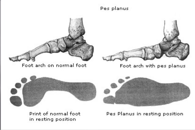
Causes
Adult acquired flatfoot is caused by inflammation and progressive weakening of the major tendon that it is responsible for supporting the arch of the foot. This condition will commonly be accompanied by swelling and pain on the inner portion of the foot and ankle. Adult acquired flatfoot is more common in women and overweight individuals. It can also be seen after an injury to the foot and ankle. If left untreated the problem may result in a vicious cycle, as the foot becomes flatter the tendon supporting the arch structure becomes weaker and more and more stretched out. As the tendon becomes weaker, the foot structure becomes progressively flatter. Early detection and treatment is key, as this condition can lead to chronic swelling and pain.
Symptoms
Initially, flatfoot deformity may not present with any symptoms. However, overtime as the tendon continues to function in an abnormal position, people with fallen arches will begin to have throbbing or sharp pain along the inside of the arch. Once the tendon and soft tissue around it elongates, there is no strengthening exercises or mechanism to shorten the tendon back to a normal position. Flatfoot can also occur in one or both feet. If the arch starts to slowly collapse in one foot and not the other, posterior tibial dysfunction (PTTD) is the most likely cause. People with flatfoot may only have pain with certain activities such as running or exercise in the early phase of PTTD. Pain may start from the arch and continue towards the inside part of the foot and ankle where the tendon courses from the leg. Redness, swelling and increased warmth may also occur. Later signs of PTTD include pain on the outside of the foot from the arch collapsing and impinging other joints. Arthritic symptoms such as painful, swollen joints in the foot and ankle may occur later as well due to the increased stress on the joints from working in an abnormal position for a long period of time.
Diagnosis
The history and physical examination are probably the most important tools the physician uses to diagnose this problem. The wear pattern on your shoes can offer some helpful clues. Muscle testing helps identify any areas of weakness or muscle impairment. This should be done in both the weight bearing and nonweight bearing positions. A very effective test is the single heel raise. You will be asked to stand on one foot and rise up on your toes. You should be able to lift your heel off the ground easily while keeping the calcaneus (heel bone) in the middle with slight inversion (turned inward). X-rays are often used to study the position, shape, and alignment of the bones in the feet and ankles. Magnetic resonance (MR) imaging is the imaging modality of choice for evaluating the posterior tibial tendon and spring ligament complex.
Non surgical Treatment
Icing and anti-inflammatory medications can reduce inflammation and physical therapy can strengthen the tibial tendon. Orthotic inserts that go inside your shoes are a common way to treat and prevent flatfoot pain. Orthotics control the position of the foot and alleviate areas of pressure. In some cases immobilization in a cast or walking boot is necessary to relieve symptoms, and in severe cases surgery may be required to repair tendon damage. 
Surgical Treatment
In cases of PTTD that have progressed substantially or have failed to improve with non-surgical treatment, surgery may be required. For some advanced cases, surgery may be the only option. Symptomatic flexible flatfoot conditions are common entities in both the adolescent and adult populations. Ligamentous laxity and equinus play a significant role in most adolescent deformities. Posterior tibial tendon dysfunction (PTTD) is the most common cause of adult acquired flatfoot. One should consider surgical treatment for patients who have failed nonoperative therapy and have advancing symptoms and deformities that significantly interfere with the functional demands of daily life. Isolated Joint Fusion. This technique is used for well reducible flat foot by limiting motion at one or two joints that are usually arthritic. The Evans Anterior Calcaneal Osteotomy. This is indicated for late stage II adult acquired flatfoot and the flexible adolescent flatfoot. This procedure will address midtarsal instability, restore the medial longitudinal arch and reduce mild hind foot valgus. The Posterior Calcaneal Displacement Osteotomy (PCDO). This technique is indicated for late stage I and early stage II PTTD with reducible Calcaneal valgus. This is often combined with a tendon transfer. A PCDO is also indicated as an adjunctive procedure in the surgical reconstruction of the severe flexible adolescent flatfoot. Soft tissue procedure. On their own these are not very effective but in conjunction with an osseous procedure, soft tissue procedures can produce good outcome. Common ones are tendon and capsular repair, tendon lengthening and transfer procedures. Flat foot correction requires lengthy post operative period and a lot of patience. Your foot may need surgery but you might simply not have the time or endurance to go through the rehab phase of this type of surgery. We will discuss these and type of procedures necessary for your surgery in length before we go further with any type of intervention.