The Way To Diagnose Posterior Calcaneal Spur

Overview
Heel spurs occur in at least half the people who have plantar fasciitis, a painful condition involving the thick tissue that runs between your heel bone and your toes. In the past, doctors often performed surgery to remove heel spurs, believing them to be the cause of the pain associated with plantar fasciitis. In treating plantar fasciitis now, doctors rely more on ice, arch supports, physical therapy and pain medications, and surgery is rarely performed.
Causes
A major cause of heel spur pain comes from the development of new fibrous tissue around the bony spur, which acts as a cushion over the area of stress. As this tissue grows, a callus forms and takes up even more space than the heel spur, leading to less space for the thick surrounding network of tendons, nerves, ligaments and supporting tissue. These important structures in the foot have limited space because of calcium or tissue buildup, which leads to swelling and redness of the foot, and a deep throbbing pain worsened with exercise.

Symptoms
Symptoms of heel spur syndrome often include pain early in the morning or after rest, as you take the first few steps. It may also include severe pain after standing or walking long hours, especially on hard cement floors. Usually more pain exist while wearing a very flat soled shoe. A higher heel may actually relieve the pain as an arch is created. The pain is usually sharp, but can also be a dull ache. The pain may only be at the bottom of the heel, or may also travel along the arch of the foot.
Diagnosis
A heel spur is often seen on X-ray as a bony protrusion, which can vary in size. However, because a Heel Spur only indicates increased load on the plantar fascia, and not pain, an ultra sound may be required to assess other actual cause of the heel pain such and may include checking to see if the plantar fascia is inflamed or degenerated.
Non Surgical Treatment
Exercise. If you think your pain is exercise-related, change your exercise routine, environment, or foot-ware, and emphasize movements and/or body parts that do not cause pain. Mind/Body. Occasionally foot pain can be related to stress. The body may respond with generalized tension that contributes to pain in many areas, including the feet. Hypnosis and guided imagery are worth exploring if an anatomical problem is not apparent. Supplements. Natural anti-inflammatories can be just as effective as ibuprofen or other over-the-counter pain relievers with fewer side effects. Try one of the following. Ginger (Zingiber officinale) standardized to 5-6% gingerols and 6% shogoals, take one to two 500 mg tablets three to four times daily. Turmeric (Curcuma longa) standardized to 95% curcuminoids, take 400-600 mg three times daily. Also, be sure that you're wearing well-fitting shoes, the proper shoes for each activity, and that you buy new foot-ware as soon as you notice signs of wear. You can also take the pressure off your heel with a donut-shaped heel cushion or a heel-raising pad placed in your shoe. Acupuncture can also relieve the pain, as can for some sufferers, magnetic shoe inserts, although the evidence behind their effectiveness is not conclusive. Osteopathic or chiropractic manipulation can help with soft tissue pain in and around the feet.
Surgical Treatment
Surgery, which is a more radical treatment, can be a permanent correction to remove the spur itself. If your doctor believes that surgery is indicated, he will recommend an operation - but only after establishing that less drastic methods of treatment are not successful.
Bursitis Of The Foot Bursal Cyst
Overview
Bursitis means inflammation of a bursa, a sac that lines many joints and allows tendons and muscles to move easily when the joint is moving. In the heel, bursitis may cause pain at the underside or back of the heel. In some cases, heel bursitis is related to structural problems of the foot that cause an abnormal gait (way of walking). In other cases, wearing shoes with poorly cushioned heels can trigger bursitis.
Causes
The swelling is the result of the blockage of blood, tissue fluids and circulation in the heel because their normal movement has been disrupted by the force of the injury. Just like cars back up behind a traffic jam, causing congestion, exhaust and overheating, blood and fluids back up behind the injured heel, causing pain, inflammation, lumps and swelling.
Symptoms
Patients with this condition typically experience pain at the back of the ankle and heel where the Achilles tendon attaches into the heel bone. Pain is typically experienced during activities requiring strong or repetitive calf contractions (often involving end of range ankle movements) such as walking (especially uphill), going up and down stairs, running, jumping or hopping (especially whilst wearing excessively tight shoes). Often pain may be worse with rest after these activities (especially that night or the following morning). The pain associated with this condition may 'warm up' with activity in the initial stages of injury. As the condition progresses, patients may experience symptoms that increase during sport or activity, affecting performance. In severe cases, patients may walk with a limp or be unable to weight bear on the affected leg. Other symptoms may include tenderness on firmly touching the affected bursa and swelling around the Achilles region.
Diagnosis
Plain radiographs of the calcaneus may reveal a Haglund deformity (increased prominence of the posterosuperior aspect of the calcaneus). However, on weight-bearing lateral radiographs, the retrocalcaneal recess often appears normal even in patients with retrocalcaneal bursitis, limiting its usefulness in making this diagnosis.Radiographs may be used as a diagnostic measure to support a clinician?s diagnosis of retrocalcaneal bursitis. Individuals with retrocalcaneal bursitis may have an absence of the normal radiolucency (ie, blunting) that is seen in the posteroinferior corner of the Kager fat pad, known as the retrocalcaneal recess or bursal wedge. This may occur with or without an associated erosion of the calcaneus.
Non Surgical Treatment
Specific treatment for bursitis will be determined by your doctor based on your age, overall health, and medical history. Extent of the condition. Your tolerance for specific medications, procedures, or therapies. Expectations for the course of the condition. Your opinion or preference. The treatment of any bursitis depends on whether or not it involves infection. Aseptic bursitis. A noninfectious condition caused by inflammation resulting from local soft-tissue trauma or strain injury. Treatment may include R.I.C.E. Rest, Ice, Compression, and Elevation. Anti-inflammatory and pain medications, such as ibuprofen or aspirin. Aspiration of the bursa fluid for evaluation in the laboratory. Injection of cortisone into the affected area. Rest. Splints.
Prevention
Prevention can be accomplished by controlling your foot structure with good supportive shoes or arch supports. Pay attention to early signs of friction like blister formation. This tells you where the areas that are more likely to cause a bursa to form and subsequently a bursitis.
Hammer Toe Fusion Implant
 Overview
A hammertoe is a misshapen second, third, or fourth toe. The toe bends up at the middle joint. The toe becomes a hammertoe because a muscle in the toe isn?t working properly or is too weak, increasing pressure on the tendons and the toe joints. Muscles normally work in twos to bend and straighten toes. If the toe stays bent too long, a hammertoe develops. Ill-fitting shoes, arthritis, heredity, even an injury, can cause the hammertoe to form. To add insult to injury, corns and calluses are common on top of hammertoes because the toe is rubbing against the shoe.
Causes
Hammer toe may also be caused by other medical conditions such as rheumatoid arthritis, osteoarthritis, or stroke because these forms of illnesses involve affectation of the person's muscles and nerves. Diabetes is also a causative factor for hammer toes due to diabetic neuropathy, which often times accompanies advanced instances of diabetes. Injury to a person's toes may also cause hammer toes, particularly if the injury involves breaking of the toes. In some instances, hammer toes may be hereditary. Some people may be genetically predisposed to develop the condition because of the natural structure of their bodies.
 Symptoms
Hammertoe and mallet toe feature an abnormal bend in the joints of one or more of your toes. Moving the affected toe may be difficult or painful. Corns and calluses can result from the toe rubbing against the inside of your shoes. See your doctor if you have persistent foot pain that affects your ability to walk properly.
Diagnosis
Your doctor is very likely to be able to diagnose your hammertoe simply by examining your foot. Even before that, he or she will probably ask about your family and personal medical history and evaluate your gait as you walk and the types of shoes you wear. You'll be asked about your symptoms, when they started and when they occur. You may also be asked to flex your toe so that your doctor can get an idea of your range of motion. He or she may order x-rays in order to better define your deformity.
Non Surgical Treatment
Your podiatrist may recommend one or more of these treatments to manage your hammer toes. Wear shoes with roomy toe boxes which don?t force your toes together. Exercises to stretch and strengthen muscles in the toes. Over the counter toe straps, cushions, and corn pads. Custom orthotic inserts. Toe caps or toe slings. In severe cases, surgery to release the muscles in the affected toes.
Surgical Treatment
There are generally two methods surgeons use to correct hammer toes, they are joint resection (arthroplasty) or bone mending (fusion), and the location where this is performed on the toe depends on where the toe is buckled. Its important to recognize that most of the surgical work involved the joints of the toe, not the joint of the ball of the foot. Sometimes a toe relocation procedure is needed when the joint of Hammer toe the ball of the foot is malaligned (subluxed or dislocated).
 Prevention
Skin creams can help maintain skin softness and pliability. A pumice stone or loofah sponge can help get rid of dead skin. Taking a warm footbath for 10 minutes two or three times a week will keep the feet relaxed and help prevent mild foot pain caused by fatigue. Adding 1/2 cup of Epsom salts increases circulation and adds other benefits. Taking footbaths only when the feet are painful is not as helpful.
Hammertoe Repair Implant
 Overview
A hammertoes is a toe that becomes permanently bent in the middle so that the end of the toe points downward. The portion of the toe before the joint where the bend occurs tends to arch upward. A hammer toe takes years to develop. Once the toe becomes permanently bent, corns or calluses may form. Treatment helps control symptoms in many people, but surgery is sometimes needed to straighten the toe.
Causes
Hereditary and shoe gear are probably the most likely reasons to develop a hammer toe. Tight pointy shoes may cause a hammer toes. High heels also can cause hammer toes. A deformed toe often develops over time, and certain types of feet may be predisposed. Some patients may develop a hammer toe or cross over toe (of the 2nd toe) due to a bunion of the big toe.
 Symptoms
Common symptoms of hammertoes include pain or irritation of the affected toe when wearing shoes. corns and calluses (a buildup of skin) on the toe, between two toes, or on the ball of the foot. Corns are caused by constant friction against the shoe. They may be soft or hard, depending upon their location. Inflammation, redness, or a burning sensation. Contracture of the toe. In more severe cases of hammertoe, open sores may form.
Diagnosis
Your doctor is very likely to be able to diagnose your hammertoe simply by examining your foot. Even before that, he or she will probably ask about your family and personal medical history and evaluate your gait as you walk and the types of shoes you wear. You'll be asked about your symptoms, when they started and when they occur. You may also be asked to flex your toe so that your doctor can get an idea of your range of motion. He or she may order x-rays in order to better define your deformity.
Non Surgical Treatment
Any forefoot problems that cause pain or discomfort should be given prompt attention. Ignoring the symptoms can aggravate the condition and lead to a breakdown of tissue, or possibly even infection. Conservative treatment of mallet toes begins with accommodating the deformity. The goal is to relieve pressure, reduce friction, and transfer forces from the sensitive areas. Shoes with a high and broad toe box (toe area) are recommended for people suffering from forefoot deformities such as mallet toes. This prevents further irritation in the toe area from developing. Other Hammer toe conservative treatment includes forefoot supports such as gel toe caps, gel toe shields and toe crests. Gel forefoot supports provide immediate comfort and relief from common forefoot disorders without drying the skin.
Surgical Treatment
In more advanced cases of hammer toe, or when the accompanying pain cannot be relieved by conservative treatment, surgery may be required. Different types of surgical procedures are performed to correct hammer toe, depending on the location and extent of the problem. Surgical treatment is generally effective for both flexible and fixed (rigid) forms of hammer toe. Recurrence following surgery may develop in persons with flexible hammer toe, particularly if they resume wearing poorly-fitted shoes after the deformity is corrected.
Pain After Tailor'S Bunionectomy
Overview
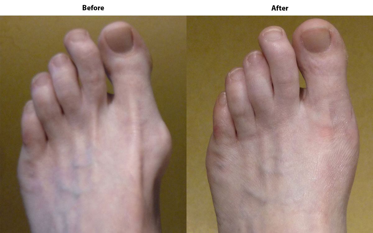
 A simple bunion is an abnormal bump of bone that is formed at the head of our old friend, the first metatarsal bone. The bunion can either be on the top or side of the first metatarsal bone. In a more advanced bunion deformity, called Hallux Abducto Valgus, there starts to be a movement of the big toe toward the second toe. The most severe bunion is when the first toe not only moves toward the second toe, but it overlaps or underlaps the second toe. Bunions can cause swelling of the feet to occur.
Causes
Bunions can be caused by improper footwear. Genetics. Foot injuries. Congenital deformities. Medical conditions such as arthritis. Stress on feet. Bunions are mainly caused by genetics. The bunion itself is not inherited, but the person?s hereditary foot type and gait pattern makes them more prone to developing bunions.You can also begin to develop bunionsby wearing shoes that are too tight or too small. When you wear shoes of this nature, your toes are squeezed together. Bunions are not caused by crowding of the toes, but wearing tight shoes can worsen the condition and cause symptoms to appear sooner. Some people are born with birth defects that put them at higher risk for developing bunions.
Symptoms
Pain or soreness. Inflamattion and redness. A burning sensation. Numbness on the side of the great toe. Other conditions which may occur secondary to bunions include calluses on the big toe, sore between the toes, ingrown toenails, and stiffness of the joint where the great toe attaches to the foot.
Diagnosis
When an x-ray of a bunion is taken, there is usually angulation between the first metatarsal bone and the bones of the big toe. There may also be angulation between the first and second metatarsal bones. These angular irregularities are the essence of most bunions. In general, surgery for bunions aims to correct such angular deformities.
Non Surgical Treatment
Padding with a number of different materials (eg felt) to reduce pressure on the painful prominence of the bunion. Physical therapy can be used to help with the symptoms and improve the range of motion (this is particularly helpful if the pain is coming from inside the joint, rather than from shoe pressure). Manipulation of the joint can be used to help with this (manipulation will never correct the alignment of the joint). Any corns and calluses that are causing symptoms should be treated. The correct fitting of footwear is essential for anyone who is serious about doing something about their bunions and hallux valgus. It may be possible to have your shoes stretched over the area of the bunion to also relieve pressure. Foot orthotics may be useful in helping with the instability about the joint. They may be more helpful if there are other symptoms in the foot as well, as their use in "treating" bunions is controversial. They may play a role in slowing progression and in the prevention of bunions developing again after surgical correction. Exercises can be important in maintaining the mobility of the joint in those with bunions, this is especially important for the arthritic type pains that may be originating from inside the joint and for the prevention of these painful symptoms in the future. 
Surgical Treatment
If other treatments don?t help and your bunion is very painful, you may be referred to an orthopaedic or a podiatric surgeon for assessment. There are over 130 different operations that can be carried out to treat bunions. The simplest operations are called bunionectomies. The majority of the operations aim to correct the alignment of your big toe. This will narrow your foot and straighten out your big toe joint as much as possible. An operation won?t return your foot back to normal, but most people find that surgery reduces their symptoms and improves the shape of their foot. The operation your surgeon will advise you to have will depend on how severe your bunion is and whether or not you have arthritis.
Prevention
The best protection against developing bunions is to protect and care for your feet every day. Avoid tight and narrow-fitting shoes. Limit your use of high heels. Wear comfortable shoes with adequate space between your longest toe and the end of the shoe. Getting treatment for very flat or very high-arched feet (if you are experiencing symptoms) will give your feet the proper support and help maintain stability and balance.
The Treatment Of Over-Pronation
Overview
Overpronation is a term that gets thrown around a lot by psuedo-experts and there is so much written online about it, that it can get very confusing to determine what is legitimate and what is not. Pronation is a normal motion of the foot that occurs when the ankle rolls inwards and the arch collapses. This is normal. Its not something evil. Overpronation is assumed to be when there is too much of it. There certainly is debate as to just how much is too much, as some people have large amounts and have no problems, other have small amounts and do have problems. It all comes down to individual differences and how much force is needed to stop the foot moving. The greater the force, regardless of the amounts of pronation, the more likely it is to be a problem. Some still like to debate if it is a problem or not. Some studies have shown that its not a problem and other studies have shown it is. If the data from all these studies are pooled, then the conclusion was that, yes, overpronation is a problem that was statistically significant, but it was only a small risk factor for problems. An associated finding of overpronation during a gait analysis is an abductory twist.
Causes
Over-pronation occurs when the foot collapses too far inward stressing the plantar fascia (the area underneath the arch of the foot.) Normally, one pronates every time he or she walks, but excessive pronation is called over-pronation. When this occurs it can cause pain in the feet, knees, hips, low back and even the shoulder. Decreasing over-pronation, which is very prominent in runners, will help add endurance, speed and efficiency to your run and ultimately place less stress on your body.
Symptoms
When standing, your heels lean inward. When standing, one or both of your knee caps turn inward. Conditions such as a flat feet or bunions may occur. You develop knee pain when you are active or involved in athletics. The knee pain slowly goes away when you rest. You abnormally wear out the soles and heels of your shoes very quickly.
Diagnosis
If you have flat feet or low arches, chances are you overpronate. Although not always the case, the lower your arches the greater the overpronate. Stand on a hard surface (in front of a mirror if you need to) and look at your feet, flat feet or low arches are easy to spot. If your feet look flatter than a pancake, have a look at your ankles and see if they seem collapsed or straight. If they are, you're overpronating.
Non Surgical Treatment
Anti-Pronation Insoles provide a unique foot support system that aligns the lower body. The major cause of foot and leg pain is over pronation (rolling over of the feet) which causes excessive pressure on the muscles, ligaments and bones of the lower body. Running insoles treat the underlying cause of over pronation and prevent future occurrences of the associated foot or leg condition. A project conducted at the NIKE Sport Research Laboratory studied the effects of orthotics on rear foot movement in running. Nine well-trained runners who wore orthotics were chosen as subjects. The results of the study indicated that orthotics reduced rear foot movement by roughly one degree or approximately nine percent of the amount found in runners not using orthotics. The average reduction of the maximum velocity of pronation was fifteen percent. Thus this study indicates that orthotics and insoles control over pronation which will treat and prevent many sporting injuries.
Prevention
Wearing the proper footwear plays a key role as a natural way to help pronation. Pronated feet need shoes that fit well, provide stability, contain supportive cushioning, are comfortable and allow enough room for your foot to move without causing pain or discomfort. Putting special inner heel wedges, known as orthotics, into your shoes can support a flatfoot while lowering risks of developing tendinitis, according to the American Academy of Orthopaedic Surgeons. More extensive cases may require specially fitted orthopaedic shoes that support the arches.
The Way To Diagnose Severs Disease?
Overview
One of the more common limitations to children enjoying their youthful energy is Sever?s Disease. Sever?s Disease is a pathology that affects the heels of children in their high growth years (usually around 9-15), and it can be quite painful. It usually occurs due to repetitive trauma or impact at the heel. This means that if your child spends a lot of time running around (in sports, or just with friends) the continuous impact of his or her heel to the ground can cause this! Usually, most children will complain of heel pain specifically when they are active; these are considered milder cases. In more severe cases the pain may never cease, whether the child is active or not.
Causes
During the growth spurt of early puberty, the bones often grow faster than the leg muscles and tendons. This can cause the muscles to become very tight and overstretched, the heel becomes less flexible and this build-up of pressure can result in redness, swelling, tenderness and pain at the heel.
Symptoms
Signs and symptoms of Sever?s disease include heel pain can be in one or both heels, and it can come and go over time. Many children walk or run with a limp, they may walk on their toes to avoid pressure on their heels. Heel pain may increase with running or jumping, wearing stiff, hard shoes (ex. soccer cleats, flip-flops) or walking barefoot. The pain may begin after increasing physical activity, such as trying a new sport or starting a new sports season.
Diagnosis
Sever disease is most often diagnosed clinically, and radiographic evaluation is believed to be unnecessary by many physicians, but if a diagnosis of calcaneal apophysitis is made without obtaining radiographs, a lesion requiring more aggressive treatment could be missed. Foot radiographs are usually normal and the radiologic identification of calcaneal apophysitis without the absence of clinical information was not reliable.
Non Surgical Treatment
The good news is that the condition doesn?t cause any long-term foot problems. Symptoms typically go away after a few months. The best treatment is simply rest. Your child will need to stop or cut down on sports until the pain gets better. When she's well enough to return to her sport, have her build up her playing time gradually. Your doctor may also recommend ice packs or nonsteroidal anti-inflammatory drugs (NSAIDs), such as ibuprofen or naproxen, to relieve the pain. Supportive shoes and inserts that reduce stress on the heel bone. These can help if your child has another foot problem that aggravates Sever?s disease, such as flat feet or high arches. Stretching and strengthening exercises, perhaps with the help of a physical therapist. In severe cases, your child may need a cast so her heel is forced to rest.
Recovery
In some cases, children will simply outgrow Sever's Disease when they reach a certain age, but this does not mean that symptoms should be ignored. If children express that they are in pain, this should always be taken seriously by their parents or guardians. Heel pain may be a sign of Sever's Disease and this condition should not be left untreated, due to the damage it can cause to the growing heel bones. Scheduling a doctor's appointment is always the first step to take in gaining a diagnosis of symptoms and speedy help for the child.